
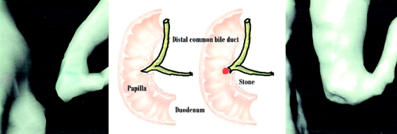
FIGURE 4. Normal (left) and pathological sphincter (right). Since MRI is less accurate for the analysis of the distal tract of CBD, the 3D reconstruction can make stone detection easier by enhancing the differences in shape of the distal tract of CBD.