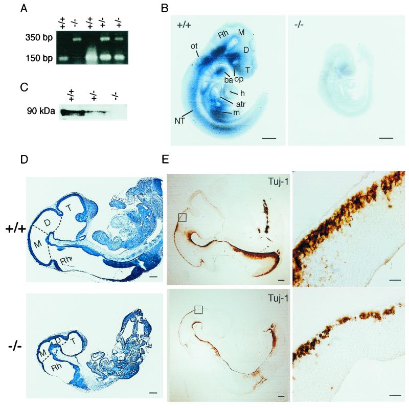
FIG. 2.
Growth retardation of Dyrk1A−/− embryos. (A) Genotyping of wild-type (Dyrk1A+/+), heterozygous (Dyrk1A+/−), and homozygous (Dyrk1A−/−) embryos by PCR analysis of DNA from embryonic yolk sac tissue. Locations of primers used for amplification of wild-type and targeted alleles are shown in Fig. 1B. (B) Whole-mount in situ hybridization in Dyrk1A+/+ and Dyrk1A−/− E9.5 embryos using a riboprobe containing the deleted Dyrk1A sequence. The absence of signal in the Dyrk1A−/− embryo and its significant size reduction compared to the wild-type embryo can be appreciated. Bars, 1 mm. (C) Western blot analysis of equivalent amounts of protein extracts from Dyrk1A+/+, Dyrk1A+/−, and Dyrk1A−/− embryos with an anti-Dyrk1A specific antibody. The typical cluster of Dyrk1A bands around 90 kDa is shown. (D) Sagittal sections of cresyl violet-stained Dyrk1A+/+ and Dyrk1A−/− embryos at E10.5. The differences between control and homozygous embryos in size and organ development, as well as in the proportions of the brain vesicles, can be appreciated. Bars, 1 mm. (E) (Left panels) Sagittal sections of E10.5 Dyrk1A+/+ and Dyrk1A−/− embryos stained with an anti-β-tubulin antibody (Tuj-1) show a significant decrease in immunostaining for the mutant embryo. Bars, 2 mm. (Right panels) Areas within squares in left panels are amplified, showing fewer immunopositive postmitotic neurons in Dyrk1A−/− embryos than in wild-type embryos. Bars, 200 μm. Abbreviations: atr, common atrial chamber; ba, branchial arch; D, diencephalon; h, heart (truncus arteriosus and bulbus cordis); M, mesencephalon; m, mesodermal tissue; NT, neural tube; op, optic vesicle; ot, otic vesicle; Rh, rhombencephalon; T, telencephalon.

