
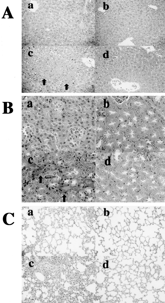
FIGURE 6. The pathologic findings of the IL-18-treated burned mice at 24 hours after the mild E. coli challenge. The liver is shown as A (hematoxylin and eosin, original magnification ×200), the kidney is B (hematoxylin and eosin, original magnification ×300) and the lung is C (hematoxylin and eosin, original magnification ×200). The nontreated burned mice (n = 4) are labeled as a, nontreated (control) unburned mice (n = 4) as b, IL-18-treated burned mice (n = 4) as c, and IFN-γ Ab + IL-18-treated burned mice (n = 4) as d, in each panel.