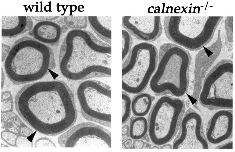
FIG. 3.
Electron microscopy on sciatic nerve fibers from wild-type and cnx−/− mice. The left panel shows a sciatic nerve fiber from a wild-type control mouse at a 22,725-fold magnification, while the right panel shows a comparable section from a calnexin gene knockout mouse. Arrowheads indicate normal myelination in both wild-type and calnexin gene-deficient mice.

