Figure 4.
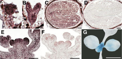
Expression Pattern of AP2.
(A) to (F) In situ hybridization using AP2 antisense probe ([A] to [C] and [E]) and sense control ([D] and [F]). Signal is detected as reddish color. Background staining independent of the in situ procedure is marked (asterisks). Globular stage embryo (A), heart stage embryo (B), mature embryo ([C] and [D]), inflorescence meristems ([E] and [F]). Bars = 20 μm in (A) and (B) and 50 μm in (C) to (F).
(G) A 14-d-old AP2:GUS seedling. Bar = 5.0 mm.

