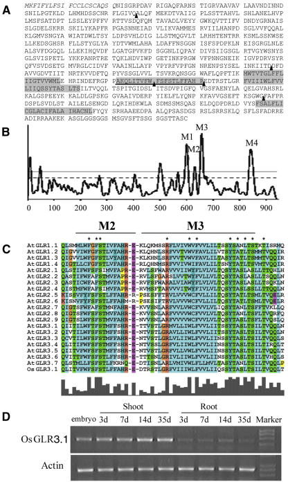
Figure 3.
Characterization of Rice GLR3.1.
(A) Deduced amino acid sequence of GLR3.1. The signal peptide is indicated in italic type; transmembrane domains (M1, M2, M3, and M4) are shaded; and the underlined region is the pore-forming domain (M2). The arrowheads indicate the positions of the exon–intron junctions identified in the genomic clone compared with the cDNA sequence. A distinctive exon–intron structure exists in the M2 domain.
(B) Hydropathy plot analysis of GLR3.1 with the program TMpred (http://www.ch.embnet.org/software/TMPRED_form.html).
(C) Alignment of the M2 and M3 domains in rice GLR3.1 with Arabidopsis GLRs. Rice GLR3.1 is shown at bottom, and the absolutely conserved residues are marked with asterisks.
(D) RT-PCR analysis of rice GLR3.1 expression at different developmental stages of rice seedlings.

