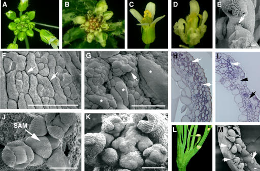
Figure 1.
Phenotypic Analyses of Wild-Type and tso2-1 Plants.
(A) A wild-type (Ler) inflorescence.
(B) A tso2-1 inflorescence showing white sectors on leaves and sepals.
(C) A wild-type flower.
(D) A tso2-1 flower showing sepals with white sectors and rough surfaces.
(E) Partial homeotic transformation in a tso2-1 floral organ, in which stigmatic tissues characteristic of carpels are found on top of an anther (arrow).
(F) Scanning electron micrograph of a wild-type sepal epidermis. Immature stomata (arrows) are interspersed with rectangular cells.
(G) A tso2-1 sepal epidermis. Clusters of epidermal cells are projected above the epidermal surface, an example of which is indicated by the arrow. Some epidermal cells are significantly large and are marked with asterisks.
(H) Cross section of a wild-type carpel valve. Chloroplasts that encircle and outline the photosynthetic mesophyll cells are stained purple and are indicated by arrows.
(I) Cross section of a tso2-1/tso2-1; rnr2b-1/+ carpel valve. Although some mesophyll cells possess chloroplasts (white arrow), others completely lack chloroplasts (black arrow) and are small. Abnormal air spaces (black arrowhead) are present beneath the epidermis.
(J) Scanning electron micrograph of a wild-type SAM showing spiral arrangements of young floral primordia.
(K) A tso2-1 SAM that is disorganized and is undergoing fasciation.
(L) A fasciated tso2-1 plant showing multiple splits of the shoot.
(M) An open tso2-1 silique showing mature seeds interspersed with aborted seeds or unfertilized ovules (arrows).
Bars = 100 μm.

