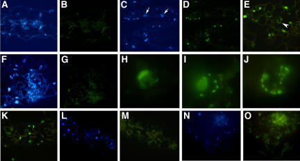
Figure 9.
PCD in tso2-1 rnr2a-1 Double Mutants Detected by the TUNEL Assay.
Images shown in (A) to (K) are from 3-week-old seedlings. Images shown in (L) and (M) and in (N) and (O) are from 6- and 11-d-old seedlings, respectively. Images in (A) to (G) and (K) to (O) are magnified ×88. Images in (H) to (J) are magnified ×366.
(A) DAPI staining of nuclei in a wild-type (Ler) leaf section.
(B) The same wild-type leaf section shown in (A) is TUNEL-negative, showing no green fluorescent signal.
(C) DAPI staining of nuclei in a tso2-1 rnr2a-1 leaf section.
(D) Many TUNEL-positive (green fluorescent) nuclei are found in the same tso2-1 rnr2a-1 leaf section shown in (C). Some nuclei (indicated by arrows in [C]) are not fluorescent, indicating that some cells have not undergone PCD.
(E) TUNEL-positive nuclei in a tso2-1 rnr2a-1 leaf section. Nuclear morphologies characteristic of different stages of PCD are shown. The arrowhead indicates a cell enlarged in (J).
(F) DAPI staining of nuclei in a tso2-1 rnr2a-1 leaf section.
(G) A negative control for the TUNEL. The same leaf section shown in (F) was not provided with terminal deoxyribonucleotidyl transferase during TUNEL labeling.
(H) A TUNEL-positive nucleus in a tso2-1 rnr2a-1 leaf section. The crescent-shaped nuclear morphology was previously described for UV-C light–treated protoplast cells undergoing PCD (Danon and Gallois, 1998).
(I) A TUNEL-positive nucleus of tso2-1 rnr2a-1 exhibiting marginalization of chromatin on the nuclear membrane and fragmentation of the nucleus into small bodies.
(J) An enlargement of the TUNEL-positive nucleus shown in (E) showing fragmented DNA bodies at the nuclear peripheral.
(K) A wild-type (Ler) leaf section treated with DNase I before detection by the TUNEL. The DNA nicks created by DNase I give positive TUNEL signals. However, none of the TUNEL-positive nuclei exhibit apoptotic characteristics, as shown in (H) to (J).
(L) DAPI staining of nuclei in a cotyledon section of a 6-d-old tso2-1 rnr2a-1 seedling.
(M) An absence of TUNEL-positive nuclei in the same cotyledon section shown in (L). Chloroplasts are visible as a result of their autofluorescence.
(N) DAPI staining of nuclei in a cotyledon section of an 11-d-old tso2-1 rnr2a-1 seedling.
(O) TUNEL-positive nuclei in the same cotyledon section shown in (N).

