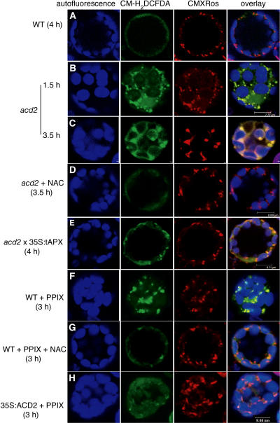
Figure 5.
ROS Production in acd2 and PPIX-Treated Protoplasts.
Protoplasts from 18-d-old wild-type, acd2 mutant, acd2 × 35S:tAPX, and 35S:ACD2 leaves were treated with or without 10 μM PPIX under light and double-stained with CM-H2DCFDA (green) and CMXRos (red). Images were observed by laser scanning confocal microscopy. Chloroplast autofluorescence (blue) was excited at 488 nm and visualized at 738 to 793 nm. This experiment was repeated three times with similar results.
(A) A wild-type control cell. Note that there were no detectable green signals after staining with CM-H2DCFDA.
(B) to (D) acd2 protoplasts were treated with or without 10 μg/mL NAC for 3.5 h. Note that the localization of the CM-H2DCFDA signals matched that of the CMXRos signals after 1.5 h. After 3.5 h, green signals were also found in the outer membrane of chloroplasts (C) and were significantly decreased by the addition of NAC (D).
(E) A cell homozygous for acd2 and 35S:tAPX.
(F) and (G) Wild-type protoplasts were treated with PPIX or PPIX + NAC for 3 h.
(H) A 35S:ACD2 cell treated with PPIX for 3 h.

