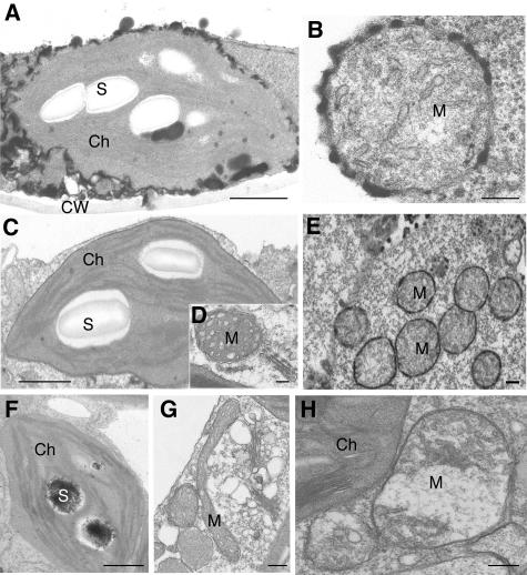
Figure 8.
Cytochemical Localization of H2O2 Using Cerium Chloride Staining of acd2 Mutant and PPIX-Treated Wild-Type Leaves.
(A) and (B) Twenty-day-old acd2 leaves with some spontaneous cell death were used. Note electron-dense deposits of cerium perhydroxidase, indicative of the presence of H2O2 in the chloroplast (A) and mitochondrial (B) outer membranes. The chloroplast and mitochondrion were structurally intact.
(C) and (D) Wild-type control leaves stained with cerium chloride.
(E) and (F) Wild-type leaves infiltrated with 50 μM PPIX for 24 h. Note the presence of H2O2 in the outer membranes of clustered mitochondria (E) and in starch grains of chloroplast (F) in cells adjacent to those that died.
(G) and (H) Protoplasts from 18-d-old wild-type leaves were treated with (H) or without (G) 10 μM PPIX for 5 h in the light and then fixed to make regular electron microscopy samples. Note that mitochondria became swollen and round in shape in a PPIX-treated cell (H) when compared with a control cell (G).
These experiments were repeated three times with similar results. Ch, chloroplast; CW, cell wall; M, mitochondrion; S, starch grain. Bars = 1 μm in (A), (C), and (F) and 200 nm in (B), (D), (E), (G), and (H).

