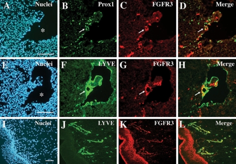
Figure 3.
FGFR-3 expression in lymphatic endothelial cells during and after embryonic development. Adjacent mouse embryo sections (E11.5) were stained for Prox1 (green) and FGFR-3 (red) (A-D) and for LYVE-1 (green) and FGFR-3 (red) (E-H). Lymphatically differentiating budding endothelial cells and resident endothelial cells in a newly formed lymphatic vessel are costained positively for Prox1 and for FGFR-3 (D). Similarly, LYVE-1-positive lymphatic endothelial cells express FGFR-3 (H). Arrows indicate a newly formed lymphatic vessel (B-D and F-H). A human neonatal foreskin section was costained for LYVE-1 and FGFR-3 (I-L). Asterisk, cardinal vein; bar, 100 μm.

