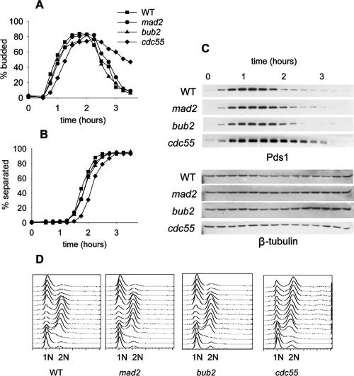
Figure 1.
Mitosis in Cdc55Δ and checkpoint mutants in a normal cell cycle. Wild-type (8715-10-2), mad2Δ (8715-6-4), bub2Δ (8579-4-4), and cdc55Δ (8766-25-4) cells were synchronized with α-factor, released into the cell cycle in YM-1 medium, and sampled at 15-min intervals. Cells were limited to a single cell cycle by addition of α-factor 100 min after release. Symbols for all graphs are as indicated in A. (A) Timing of bud emergence, determined by phase contrast microscopy. (B) Timing of sister chromatid separation at LEU2, determined by GFP fluorescence microscopy. (C) Immunoblots of PDS1-13Myc levels to assay the onset of anaphase. (D) Flow cytometry of DNA content to determine the kinetics of replication and cell division.

