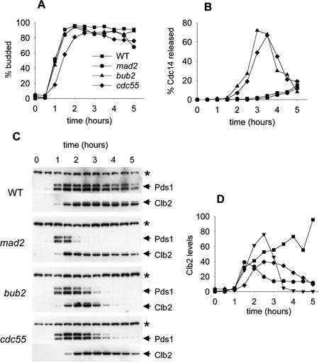
Figure 3.
Exit from mitosis in Cdc55Δ and checkpoint mutants in nocodazole. Wild-type (8905-4-3), mad2Δ (8905-1-3), bub2Δ (8906-1-3), and cdc55Δ (8866-30-2) cells were synchronized with α-factor, released into YM-1 medium containing 12 μg/ml nocodazole, and sampled at 30-min intervals for a total of 5 h. Cells were limited to a single cell cycle by addition of α-factor 100 min after release. Symbols for all graphs are as indicated in A. (A) Bud morphology, determined by phase contrast microscopy. (B) Timing of Cdc14-13Myc release from the nucleolus, determined by immunofluorescence microscopy. Each time point contains data from at least 100 cells. (C) Immunoblots to assay the stability of PDS1-3HA and Clb2-3FLAG proteins. (D) Quantitation of Clb2 levels from scanned immunoblots.

