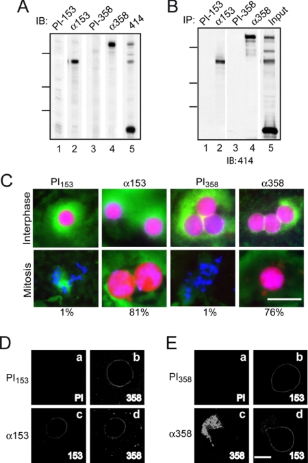
Figure 3.
Both Nup358 and Nup153 play a role in nuclear envelope breakdown. (A) Immunoblot of fractionated egg extract probed with antibody against Nup153 (lane 2) and its matched preimmune (PI153; lane 1), Nup358 antiserum (lane 4) and its matched preimmune (PI358; lane 3), and 414-reactive Nups (lane 5). (B) Immunoprecipitation of egg extract with antibodies against Nup153 (lane 2) and its matched preimmune (lane 1), Nup358 antiserum (lane 4) and its matched preimmune (lane 3). Lane 5 was loaded with the equivalent of ∼20% input. The blot was probed with mAb414. For both A and B, molecular-weight markers indicated are 214, 118, and 92 kDa. (C) Antibodies against Nup153 or its matched preimmune (0.25 μg), or Nup358 antiserum or its matched preimmune (0.5 μl) was added 15 min before the beginning of the assembly/disassembly assay. Interphase samples were taken 90 min after assembly, and mitotic samples were taken 75 min after cyclin addition. Nuclear import cargo, DNA, and membrane are shown. The percentage of intact nuclei at mitosis is indicated below the panel. Bar, 50 μm. (D) In vitro nuclei were formed after a 15-min preincubation with preimmune antibody or antibody against Nup153. The added antibody was detected by indirect immunofluorescence with goat anti-rabbit secondary antibody (a and c). The localization of Nup358 was determined by incubating with primary and secondary antibodies postfixation (b and d). (E) In vitro nuclei were formed after a 15-min preincubation with preimmune or Nup358 antiserum. The added antibody was detected by indirect immunofluorescence with a goat anti-guinea pig secondary (a and c). The localization of Nup153 was determined by incubating with primary and secondary antibodies postfixation (b and d). Bar (D and E), 20 μm.

