FIG. 6.
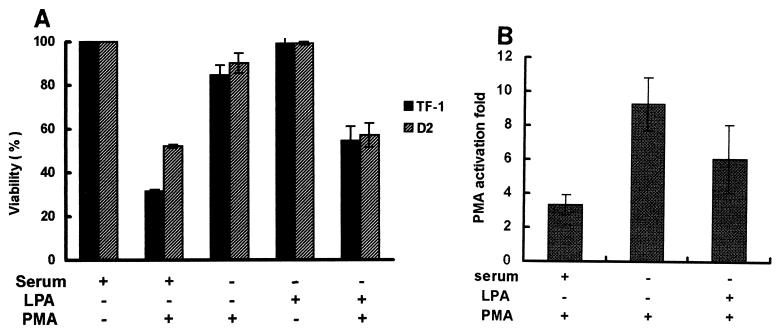
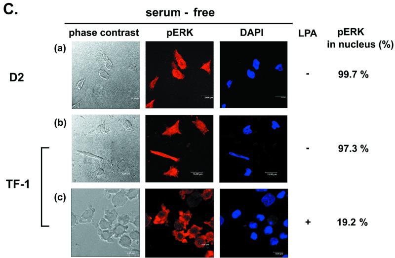
Serum and LPA can affect activation of the p21Cip1/Waf1 promoter and phospho-ERK nuclear translocation during PMA treatment. (A) TF-1 and D2 cells (2 × 10⁵/ml) were suspended in serum-free medium. After incubation of cells without or with 10% heat-inactivated fetal bovine serum or LPA (10 μM), as indicated, for 10 min, PMA (32 nM) was added to the medium. After 12 h of PMA treatment, viable cells adhering to the plates were counted. (B) D2 cells were transfected with the pWWP-Luc plasmid. After transfection for 2 days, cells were washed and suspended in serum-containing, LPA-containing, or serum-free medium, followed by treatment with PMA (32 nM) or no treatment for 3 h for luciferase assays. PMA-induced activation was calculated as described in the legend to Fig. 2B. For the cells treated with PMA in serum-containing medium, the suspended and attached cells were pooled for the luciferase activity assay. The data represent averages for three independent experiments. (C) D2 and TF-1 cells treated with PMA in serum-free medium for 3 h were fixed for immunofluorescent detection of phospho-ERK and nuclear DAPI staining (a and b). TF-1 cells incubated with LPA (10 μM) in serum-free medium were treated with PMA for 3 h for immunofluorescent detection as described above (c). The percentage of nuclear phospho-ERK-containing cells in the total number of phospho-ERK-positive cells was calculated as described in the legend to Fig. 4.


