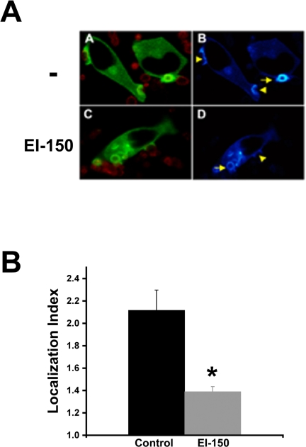
Figure 4.
DAG antagonist decreases protein kinase C-ε localization. GFP-protein kinase C-ε-expressing cells were preincubated with EI-150 (100 μM; 45 min) followed by BIgG phagocytosis. (A) GFP localization was followed by real-time confocal microscopy in control (-) or EI-150-treated cells (A and C, merge image; B and D, pseudocolor for GFP expression). Accumulation can be seen in forming phagosomes (arrowhead) and after target internalization (arrow). (B) Quantitation of GFP protein kinase C-ε localization in EI-150-treated cells. The Localization Index was calculated for phagosomes lying just beneath the plasma membrane (e.g., targets indicated by arrow in left panels). Data are presented as mean ± SEM (n > 35 events from four independent experiments). *p < 0.003 compared with untreated protein kinase C-ε-expressing cells.

