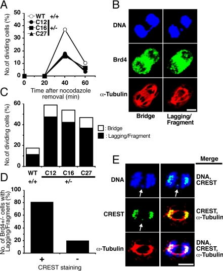
Figure 3.
Brd4+/- cells are haploinsufficient in mitotic progression after nocodazole removal. (A) Impaired anaphase progression in Brd4+/- cells after nocodazole withdrawal. Brd4+/+ and +/- cells were treated with nocodazole (100 ng/ml) for 8 h. Mitotic cells were then allowed to proceed in fresh media for indicated times (minutes) and stained for Brd4 and DNA. Cells carrying segregating chromosomes were scored as those that progressed to anaphase/telophase. A representative of three independent tests is shown. At least 250 cells were examined in each experiment. (B) Abnormal chromosomal segregation in Brd4+/- cells. Segregating chromosomes were detected by visualizing stained DNA. An example of chromosomal bridge (Bridge) and misdistributed chromosome (Lagging/Fragment) is shown. Note that Brd4 remained unloaded in both cases. Bar, 5 μm. (C) The percentage of dividing cells carrying abnormal chromosomal segregation is shown. A representative of three independent tests is shown. More than 200 anaphase/telophase cells were inspected in each experiment. (D) The percentage of dividing Brd4 +/- cells showing CREST signals on misdistributed chromosomes is shown. More than 200 anaphase/telophase cells were inspected. (E) Brd4+/- cells incubated for 40 min after release from nocodazole treatment (100 ng/ml; 8 h) were fixed and immunostained with antibody for CREST (green) and α-tubulin (red) and counterstained for DNA (blue). CREST staining of a lagging chromosome is marked by arrow. Bar, 5 μm.

