
Figure 5.
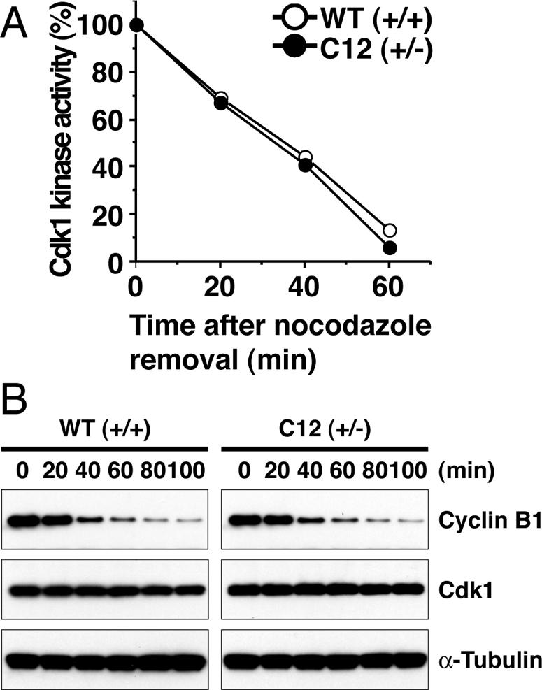
Comparable Cdk1 kinase activity and cyclin B degradation in Brd4+/+ and +/- cells. (A) Cdk1 activity was tested with histone H1 as a substrate using precipitates of lysates (30 μg) from Brd4+/+ and +/- cells that had been treated with nocodazole at 100 ng/ml for 8 h and incubated in fresh media for indicated times (minutes). Values represent the average of three samples. (B) Lysates in A were subjected to immunoblot analysis to detect cyclin B1 and Cdk1 expression. α-Tubulin was immunoblotted as a control.