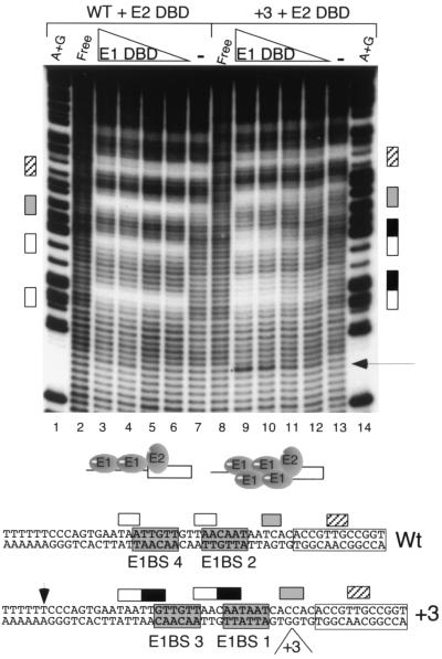
FIG. 3.
Hydroxyl radical footprinting on wt and +3 templates. Footprinting was performed on the top strand of the wt (lanes 1 to 8) and the +3 (lanes 9 to 14) ori probes in the presence of E1 and E2 DBDs as indicated at the top. E1 DBD (0.8, 0.4, 0.2, and 0.1 μg) and E2 DBD (0.7 ng) were added in lanes 3 to 6 and 9 to 12. Lanes 7 and 13 contained 70 ng of E2 DBD alone. Below, the protections are shown projected onto the DNA sequence. Hatched boxes, protections generated by E2; open boxes, protections generated by the binding of two molecules of E1 DBD to the wt probe; black boxes, extensions of these protections observed on the +3 probe; gray boxes, protections shared between E1 and E2 DBDs. Arrow, hypersensitive site that appears upon the binding of four molecules of E1 DBD. Lanes A+G and Free are as defined for Fig. 2A.

