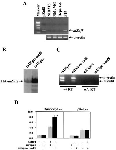
FIG. 7.
Involvement of endogenous mZnf8 in BMP signaling pathway. (A) To identify cell lines expressing endogenous mZnf8, RT-PCR was performed as described in Materials and Methods. The template in the PCR of pZnf8 is a plasmid containing the full-length mZnf8 cDNA. M refers to the 1kb+ DNA molecular size markers, purchased from Life Technologies. As a positive control, β-actin was detected in all lines. No signal was detected in no-RT controls (data not shown). (B) HA-mZnf8 was cotransfected with mU6pro-znf8 or with vector alone (mU6pro) into P19 cells. Total cell protein (20 μg) was analyzed by SDS-PAGE, and Western analysis was performed with an HA antibody. (C) P19 cells were transfected with either the vector alone (mU6pro) or mU6pro-znf8, and RT-PCR analysis was performed with mZnf8 and β-actin primers added to the same tube. Much less mZnf8 was amplified from cells transfected with mU6pro-znf8 compared to cells transfected with vector alone. (D) The 12(GCCG)-lux or pTlx-lux reporter (0.2 μg) was cotransfected with mU6pro or mU6pro-znf8 (1.0 μg) into P19 cells. Cells were treated with 100 ng of hBMP4 or bovine serum albumin per ml alone for 14 to 16 h, and luciferase assays were performed as described in Materials and Methods. Results are expressed as fold induction. The activity of cells with vector alone and without hBMP4 stimulation was defined as 1.0. Data were averaged from three independent cultures, with error bars indicating standard deviations. *, significant increase (P < 0.05).

