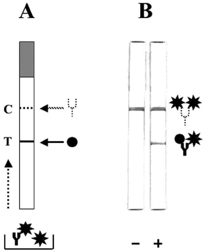
FIG. 5.

Design and use of the rapid strip assay to detect antidengue antibodies using the r-DME-M protein. (A) Schematic representation of the strip test template. The strip consists of a nitrocellulose membrane mounted on a plastic support. The shaded portion at the top represents the absorbent pad. The test line (T, indicated by the solid line) denotes the position on the strip at which the r-DME-M protein (indicated by the black circle) is immobilized as a thin line. The control line (C, indicated by the dashed line) denotes the position on the strip at which murine anti-r-DME-M polyclonal serum (indicated by the dotted Y) is coated as a thin line. Shown below is the sample receptacle containing the test serum mixed with gold-labeled r-DME-M (indicated by the black star). Antidengue IgM antibody (indicated by the solid Y) if present will form a complex with the gold-labeled r-DME-M in the sample well. The dotted arrow at the left side of the strip template indicates the direction of sample migration during the test. (B) Depiction of the actual results of the strip test performed with sera that lack (−) or contain (+) antidengue IgM antibodies. A schematic representation of the complexes formed at the T and C lines is shown to the right.
