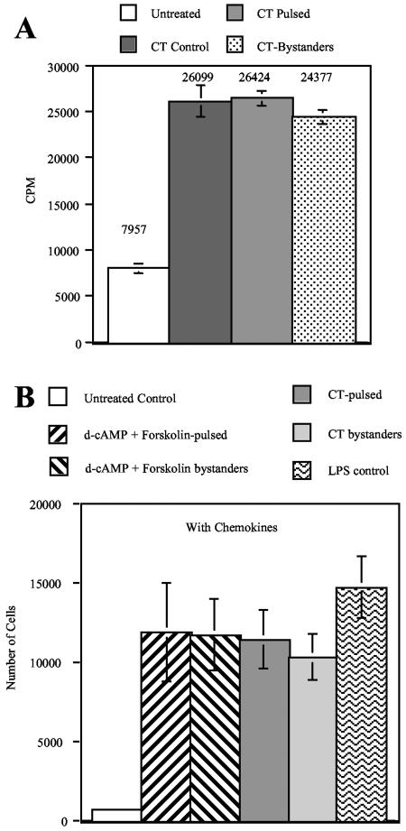
FIG. 2.
Bystander MDDC are functionally mature. (A) Bystander MDDC induce robust T-cell proliferation. MDDC prepared in Fig. 1D were used in an MLR as described in Materials and Methods. Data shown are the means and standard errors from cells derived from three different donors. (B) Migration of MDDC in response to MIP-3β and 6Ckine. MDDC (1 × 10⁵ cells) were placed into the upper chamber of the transwell system. The lower chamber contained medium supplemented with 100 ng/ml each of MIP-3β and 6Ckine. After 2 h of incubation at 37°C, the cells in the lower chamber were counted. Control cultures were kept in the absence of chemokines to assess random migration activity. Data shown are the means and standard errors of the means from cells derived from three different donors.

