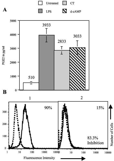
FIG. 4.
PGE2 production by MDDC. (A) MDDC were treated with 1 μg/ml CT or LPS or 1 mM d-cAMP. Twelve hours later, supernatants were removed, and PGE2 concentrations in the supernatants were measured by EIA. Data were obtained from three experiments performed on MDDC derived from different donors. The means and standard errors of the means are shown. (B) Example calculation of percent inhibition. The formula used to calculate percent inhibition is [(X − Y)/X] × 100, where X is the fraction of cells that increased the expression of a marker in the absence of the inhibitor and Y is the fraction of cells that increased the expression of a marker in the presence of the inhibitor. Panel 1 shows CD83 expression in the absence of an inhibitor, and panel 2 shows CD83 expression in the presence of an inhibitor. X is 90, Y is 15, and the equation [(90 − 15) × 100]/90 equals 83.3% inhibition.

