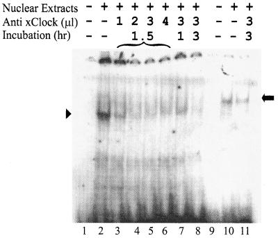
FIG. 3.
EMSAs in the presence of the xCLOCK antibody. Shifted complexes are compared after the use of the PE (lanes 1 to 8) and NE (lanes 9 to 11) as probes. The amounts of antibody and the incubation periods are labeled at the tops of the lanes. Lanes 1 and 9 are the free probes without nuclear extracts. The arrowhead and arrow mark the positions of the specific DNA-protein complexs with the PE and NE probes, respectively.

