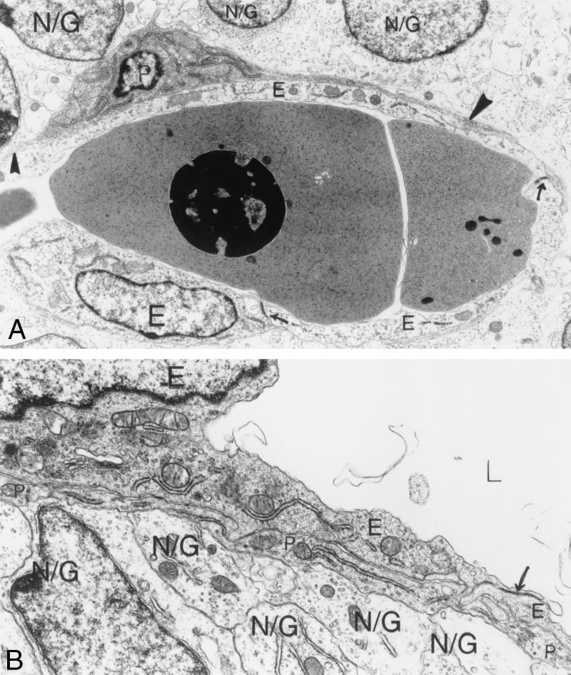
FIG. 5.
Ultrastructural analysis of E12.5 αv+/− cerebral microvessels. (A) Vascular lumen seen in longitudinal section, containing nucleated red blood cells. Normal endothelial cells (E) with closed junctions (arrows) line the lumen. One pericyte (P) has two long, narrow processes in view (solid arrowheads). Overlying brain parenchymal cells are in contact with the pericyte beneath. (B) Another view of an αv+/− cerebral microvessel shows an empty vascular lumen, two normal endothelial cells (E) connected by a closed interendothelial junction (arrow). A pericyte process (P) covers the two endothelial cells. Brain parenchymal cells and their processes (N/G) cover the overlying pericyte process completely, with little to no intervening space. Magnification: (A) 8,000×; (B) 15,500×. Abbreviations: E, endothelial cells; P, pericytes; N/G, brain parenchymal cells.

