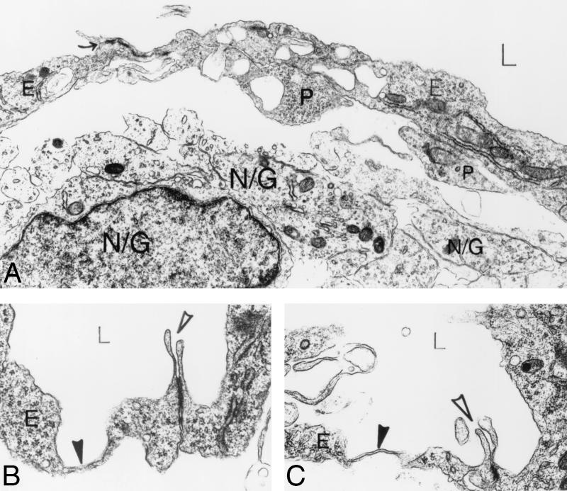
FIG. 7.
Ultrastructural analysis of αv−/− cerebral microvessels. (A) A space separates the vessel from the underlying brain parenchymal cells (N/G). Several pericyte processes (P) contact endothelial cells (E), which line the lumen (L) of the vessel. Large membrane-bound vacuoles are seen within endothelial cells and pericytes. An interendothelial cell junction is closed (arrow). Panels B and C show focal (B) and flat (C) parajunctional thinned endothelial cell cytoplasm (solid arrowheads). Note that the interendothelial cell junctions (open arrowheads) are normal, guarded by endothelial cell flaps, and are tightly closed. Magnification: (A) 10,000×; (B and C) 24,000×. Abbreviations: E, endothelial cells; P, pericytes; N/G, brain parenchymal cells.

