
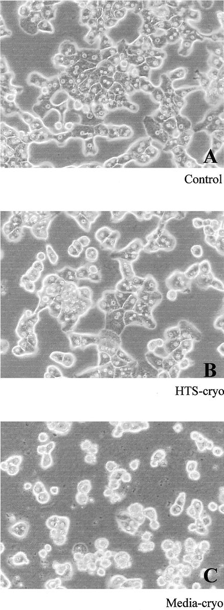
FIGURE 3. Phase contrast micrographs of hepatocytes following 14 days of culture. Cells were cultured in a collagen gel 3-dimensional configuration for 14 days. Cells cryopreserved in (B) HTS + 10% DMSO exhibited morphology similar to (A) controls (intact, hexagonal cells), whereas (C) media + 10% DMSO–cryopreserved cells were morphologically different, characterized by cellular disintegration and irregular cell shape.