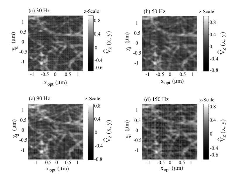
Figure 6.

Experimental AFM images of collagen (type I) obtained when using the inversion-based feedback/feedforward approach at scan frequencies of: (a) 30 Hz; (b) 50 Hz; (c) 90 Hz; and (d) 150 Hz. The scan size is 2.4 μm × 2.4 μm, and the z-Scale represents the normalized AFM-probe deflection, given by Eq. (13).
