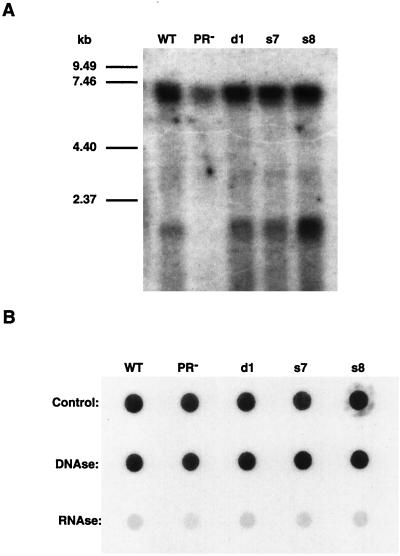
FIG. 7.
(A) RNA blot of VLP-extracted nucleic acids. The amount of VLP sample used was normalized via anti-Gag immunoblots. The nucleic acids were separated on a 1% agarose-formaldehyde gel, transferred to a GeneScreen membrane, and probed as for Fig. 4. (B) Dot blot of VLP-extracted nucleic acids. Nucleic acids from the same extraction were divided into three pools and treated with DNase or RNase A or were not treated and were probed as described above. WT, wild type.

