FIG. 1.
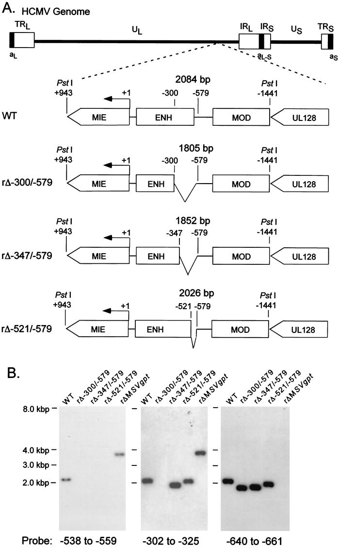
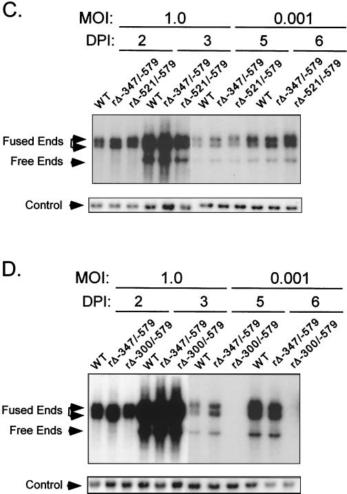
Effects on viral DNA replication of successively larger 5′-subsegmental distal enhancer deletions. (A) Schematic diagram of deleted distal enhancer segments in recombinant HCMVs rΔ-300/-579, rΔ-347/-579, and rΔ-521/-579. Base positions of deletions and PstI sites are depicted relative to the +1 RNA start-site of the MIE promoter. Predicted sizes of PstI RFLPs are shown. MIE, MIE transcription unit; ENH, enhancer; MOD, modulator; UL128, putative UL128 gene. (B) Southern blot analyses of PstI RFLPs of WT, rΔ-300/-579, rΔ-347/-579, rΔ-521/-579, and rΔMSVgpt. rΔ-300/-579, rΔ-347/-579, and rΔ-521/-579 were derived from rΔMSVgpt. Probe coordinates are given in base positions relative to the RNA start-site of the MIE promoter. (C) Abundances of WT, rΔ-347/-579, and rΔ-521/-579 genomes in HFF cells at MOI of 1.0 and 0.001. Infections were performed in parallel with equivalent input viral titers (see Materials and Methods). On the indicated day p.i. (DPI), infected-cell DNA was isolated, digested with HindIII, fractionated by gel electrophoresis, and subjected to Southern blot analysis. The 32P-labeled probe hybridizes to HCMV genomic fragments containing either the terminal repeat-long (TRL) or the internal repeat-long (IRL) region (23). The IRL is fused (Fused End) to the short genome segment, which is contained in the 17.2- and 13-kb fragments. The TRL is not fused (Free End) to the short genome segment and is contained in the 9.7-kb fragment. The blot was stripped and rehybridized to a λ-specific, 32P-labeled probe for detection of the lambda DNA internal control (Control). (D) Abundances of WT, rΔ-347/-579, and rΔ-300/-579 genomes in HFF cells at MOI of 1.0 and 0.001. The analysis was performed as described for Fig. 1C.


