
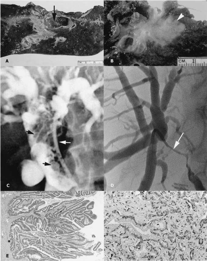
FIGURE 1. Cholangiographic, gross, and microscopic appearance of a papillary cholangiocarcinoma (left, panels A, C, E) and a nodular-sclerosing tumor (right, panels B, D, F). Note the papillary tumor within the bile-duct lumen (panel A, arrow) and the nodular-sclerosing tumor invading the hepatic parenchyma (panel B, arrow). Transhepatic cholangiogram of a papillary tumor (panel C) showing multiple filling defects that expand the duct (black arrows; the biliary drainage catheter is indicated by the white arrow). This is in contrast to the cholangiographic features of nodular-sclerosing tumors characterized by an irregular stricture that constricts the duct lumen (panel D, arrow); a transhepatic catheter is seen traversing the stricture. Histologic section of a papillary cholangiocarcinoma with no invasive component (panel E, asterisk) and an invasive nodular-sclerosing tumor associated with a desmoplastic stroma (panel F).