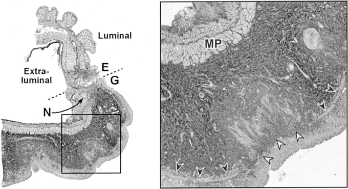
FIGURE 10. Representative trichrome-stained section of the anastomosis in an Immediate animal. Broken line represents anastomosis between the esophagus (E) and the gastric tube (G). N denotes a needle hole. The inset depicts a higher magnification to show structures of interest: muscularis propria (MP), intact muscularis mucosa (black arrowheads), and areas of atrophy in the muscularis mucosa (white arrowheads).
