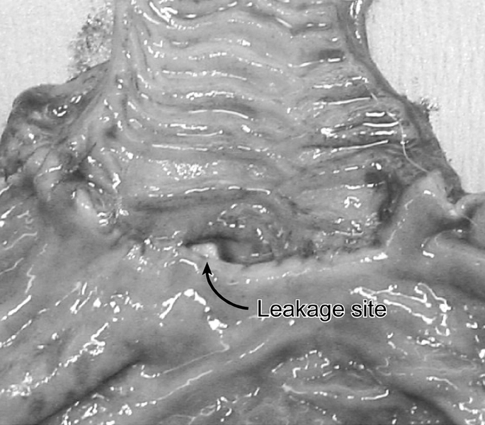
FIGURE 6. Anastomotic leakage at the esophagogastrostomy. This animal, which had been assigned to the Immediate group, developed sepsis and expired. The anastomosis was harvested, and the luminal side of the longitudinally opened specimen is displayed. The site of leakage is indicated between the esophageal (top) and gastric (bottom) conduit.
