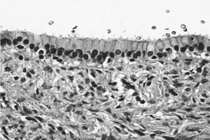
FIGURE 2. Biliary cystadenoma (40× magnification, hematoxylin and eosin stain). Lumen, upper third of frame. The mucosal surface is a simple, orderly, biliary-type columnar epithelium with basally oriented uniform and round nuclei. The mesenchymal stroma is typical of biliary cystadenomas, showing a benign spindle-cell population (“ovarian-like” stroma).
