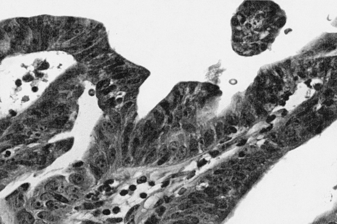
FIGURE 3. Biliary cystadenocarcinoma (40× magnification, hematoxylin and eosin stain). Increased nuclear pleomorphism and chromatin irregularity with increased epithelial cell stratification and tubulopapillary growth as compared with the biliary cystadenoma.
