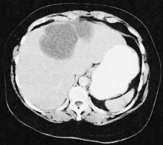
FIGURE 4. Representative CT scan image from a patient with a large biliary cystadenoma of the left lobe of the liver. Several septations are present. The interior illustrates a homogeneous appearance without studding of the internal lining sometimes seen with cystadenocarcinomas.
