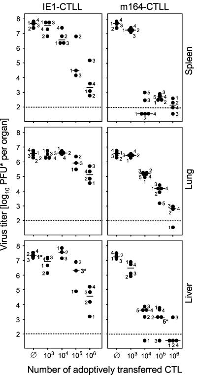
FIG. 5.
In vivo antiviral function of CTLLs. Graded numbers of CTLs were transferred intravenously into BALB/c recipients under lethal conditions of infection (6.5 Gy of total-body gamma irradiation followed by intraplantar infection with 105 PFU of mCMV). Ø, no adoptive cell transfer. Virus titers in homogenates of spleen, lung, and liver were determined on day 12 after infection. The virus plaque assay was performed under conditions of centrifugal enhancement of infectivity. Accordingly, titers of infectious virus are expressed as PFU*. Dots represent virus titers in numbered individual mice. Asterisks at the numerals mark individual mice for which the liver histopathology is documented in Fig. 6. The median values are marked by horizontal bars. The dotted line indicates the detection limit of the plaque assay.

