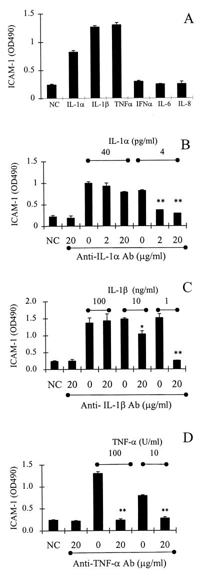
FIG. 3.
ICAM-1 expression on vascular endothelial cells is activated by recombinant cytokines. (A) ICAM-1 expression on endothelial cells incubated for 18 h with IL-1α (4 pg/ml), IL-1β (100 pg/ml), TNF-α (100 U/ml), IL-6 (1 μg/ml), and IL-8 (1 μg/ml). (B) ICAM-1 expression on endothelial cells incubated for 18 h with IL-1α in the presence or absence of anti-IL-1α antibody at the indicated concentration. (C) ICAM-1 expression on endothelial cells incubated for 18 h with IL-1β in the presence or absence of anti-IL-1β antibody at the indicated concentration. (D) ICAM-1 expression on endothelial cells incubated for 18 h with TNF-α in the presence or absence of anti-TNF-α antibody at the indicated concentration. NC, negative control (medium alone); PC, positive control (vascular endothelial cells treated for 18 h with TNF-α [10 U/ml]). Data are from three to six separate experiments with triplicate samples and are represented as the means ± standard deviations of the means. Significant differences between antibody-treated and untreated samples are indicated by * (P < 0.05) and ** (P < 0.01).

