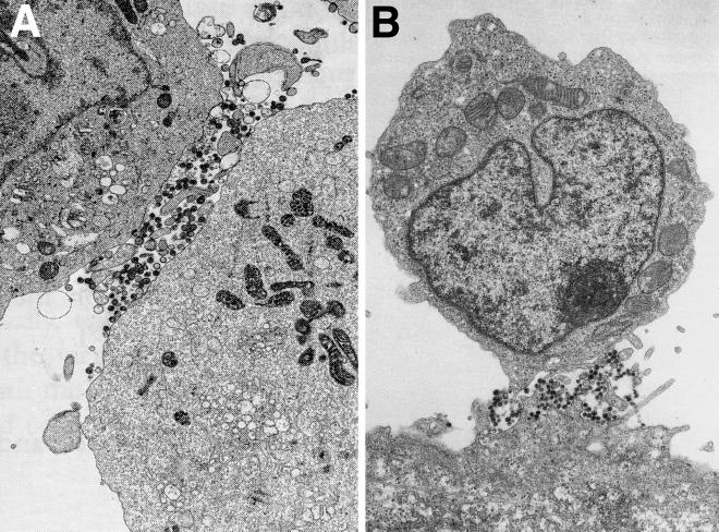
FIG. 1.
Polarized egress of HIV particles on T lymphocytes. (A) HIV-infected C8166 cells, a CD4+ T-cell line, were analyzed by electron microscopy. Virions were found preferentially at sites of cell contact. (Reprinted from AIDS [26] with permission of the publisher.) (B) HIV-infected CD4+ T cells (upper cell) adhering to a BeWo epithelial cell in the lower part of the panel. Virions were observed at sites of cell-cell contact, between microvilli extending from both cells. (Reprinted from AIDS [65] with permission of the publisher.)

