
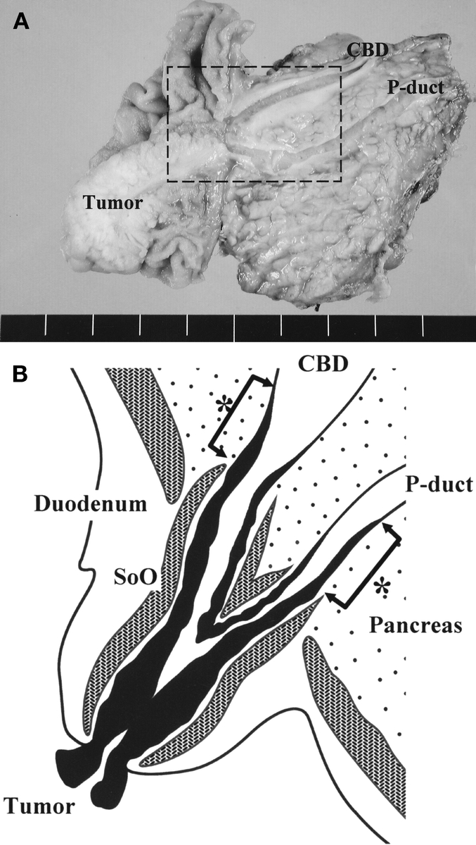
FIGURE 1. A, Surgical specimen after PD, which was resected for CBD and P-duct visualization at the same cross section. B, Schematic drawing of the rectangular area in A, showing mucosal tumor infiltration into the CBD and the P-duct of early ampullary cancer. One expert pathologist measured the length from the proximal end of the sphincter of Oddi to the upper limit of the mucosal tumor (*arrows).