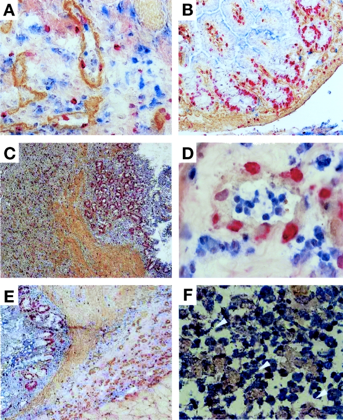
FIGURE 2. Regeneration process of rat newborn intestine. Either fresh (A–C) or 30-day-cryopreserved (D–F) newborn intestines of RT1c-7.1 rat were transplanted into RT1c-7.2 congenic rats, and specimens were multiply stained with anti-RT1c-7.2 (HIS41) mAb (blue), antitype IV-collagen mAb (brown), and anti-BrdU mAb (red). A, Blood vessels in the submucosa at 2 POD (original magnification ×100). The basement membrane was stained with antitype IV collagen mAb (brown). B, Crypts of the graft at 3 POD (original magnification ×40). C, The mucosal, submucosal and muscular layers developed at 14 POD, and numerous BrdU-positive cells were found in the crypts (original magnification ×40). D, Blood vessels in the submucosa of the cryopreserved graft at 2 POD (original magnification ×100). E, The mucosal, submucosal and muscular layers developed at 11 POD. F, Arrowheads indicate HIS41- and BrdU-double cells in the submucosal layer at 11 POD (original magnification ×100). Experiments were performed 2 times with similar results.
