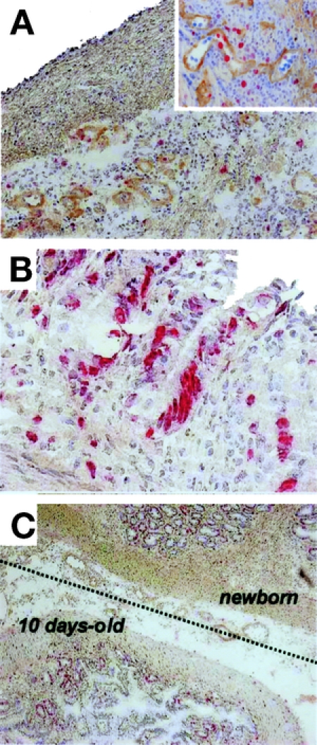
FIGURE 4. Regeneration process of 10-day-old intestinal graft in the presence of newborn graft. A fresh 10-day-old intestinal graft was transplanted subcutaneously as a twin graft in the presence of a newborn intestine. Specimens of 10-day-old grafts were multiply stained with antitype IV collagen mAb (brown) and anti-BrdU mAb (red). The counterstaining is hematoxylin. A, BrdU-positive cells were distributed in the submucosa at 2 POD (original magnification ×20). The upper-right corner inset represents a close-up view of blood vessels (original magnification ×100). B, BrdU-positive cells in the crypts at 3 POD (original magnification ×100). C, Lower view of 10-day-old intestinal graft at 10 POD (original magnification ×20). The dotted line represents the border between 10-day-old and newborn grafts.
