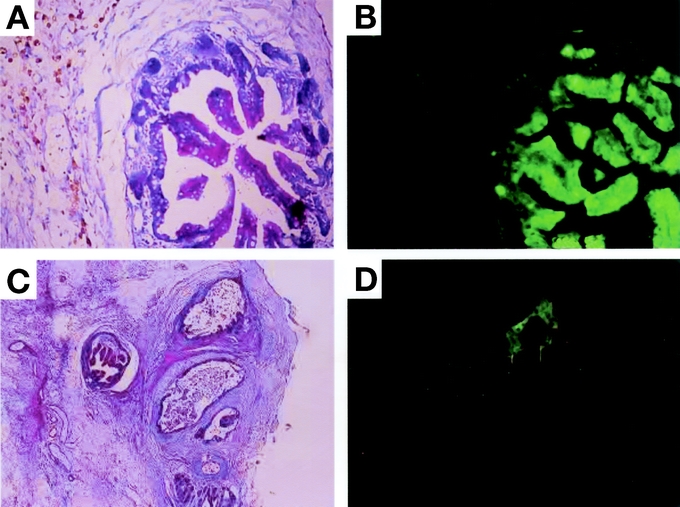
FIGURE 5. Histologic reconstitution of a 10-day-old graft from tissue aggregates. A, Neonatal intestinal grafts from GFP-transgenic Lewis rats were chopped at random and transplanted into the subcutaneous space of wild-type Lewis rats. A representative specimen is shown at 14 POD (original magnification H&E, ×40). B, GFP expression of the specimen in a (under a 489-nm excitation light, original magnification ×40). C, 10-day-old intestinal grafts from GFP-transgenic Lewis rats were chopped at random and transplanted subcutaneously together with similarly chopped newborn grafts from wild-type Lewis rats. A representative specimen is shown at 14 POD (H&E, original magnification ×20). D, GFP expression of the specimen in c (under a 489-nm excitation light, original magnification ×20). Note the substantial expression of GFP from the 10-day-old intestinal graft. One of 2 independent experiments with similar results.
