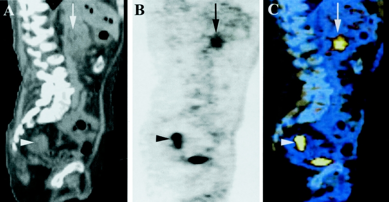
FIGURE 3. Sagittal CT (A), PET (B), and coregistered PET/CT (C) images of a 76-year-old male patient. The images show FDG-positive cancer in the head of the pancreas (arrow). In addition, FDG uptake was detected in the upper rectum (arrowhead), which referred to synchronous rectal cancer.
