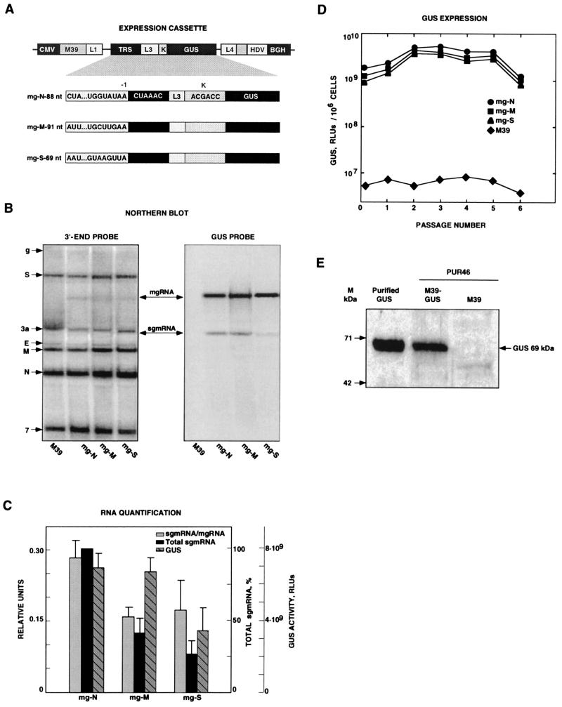
FIG. 1.
Analysis of GUS gene transcription and expression by using minigenomes that contain different virus-derived 5′ TRSs. (A) Schematic structure of expression modules based on TGEV-derived minigenome M39 cloned under the control of the CMV immediate-early promoter (CMV). TRSs include 5′-flanking sequences derived from the TRSs of the major structural genes (N, nucleocapsid gene; M, membrane gene; S, spike gene) and the CS (5′-CUAAAC-3′). The expression cassettes located below the shaded area are flanked by L1 and L4 polylinkers. This cassette includes the TRS, an insertion site (L3), an optimized Kozak sequence (K), and the GUS gene. HDV, HDV ribozyme; BGH, BGH termination and polyadenylation signals. The designations to the left of the expression cassettes indicate the origin of each TRS and the number of nucleotides inserted. (B) Northern blot analysis of intracellular RNAs extracted at passage 2 from minigenome-transfected and TGEV-infected cells. The left and right panels show hybridizations with 3′ UTR- and GUS-specific probes, respectively. The positions of the genomic RNA (g) and mRNAs (S, 3a, E, M, N, and 7) from the helper virus are indicated to the left by their acronyms. M39 RNA overlaps mRNA 3a (first lane). mgRNAs encoding the expression cassettes with different TRSs and the mRNA for the GUS gene (sgmRNA) are indicated with arrows. (C) Quantification of RNAs detected by Northern blotting. RLUs, relative luminometric units. The data shown are the means and standard errors for three independent experiments. (D) GUS activity (per 106 cells) expressed by minigenomes encoding the GUS gene under the control of TRSs derived from the N, M, and S viral genes through six passages after transfection. Background levels are those corresponding to minigenome M39, without an insert. The data shown are averages for at least three experiments with similar results. (E) Western blot analysis of GUS expression by using TGEV-derived minigenomes. Detection of the heterologous GUS protein (69 kDa) expressed by TGEV-derived minigenome M39 (M39-GUS) was performed by Western blot analysis under reducing conditions with a GUS-specific polyclonal rabbit antiserum. Molecular masses (M) are indicated on the left. Purified GUS was used as the positive control. M39-GUS, extracts from ST cells transfected with DNA coding for minigenome mg-N-88-L2 and infected with the helper virus (PUR46-MAD), obtained at passage 4. M39, extracts from ST cells transfected with minigenome M39 and infected with the helper virus.

