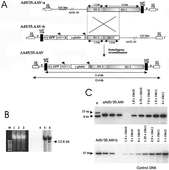
FIG. 1.
Generation of capsid-modified ΔAd.AAV vectors containing a 11.6-kb transgene γ-globin cassette. (A) Scheme for formation of ΔAd5/35.AAV genomes. The expression cassette for human γ-globin included the β promoter from −127 to the β initiation codon, which was connected in frame with the γ coding region partially deleted for intron 2 (11). Transcription of the γ-globin gene is terminated by the endogenous globin polyadenylation signal. The β promoter is linked to the 5.3-kb LCR fragment composed of HS2 and HS3 regions (6). The vector also contained the eGFP gene under the control of the MSCV promoter, which has been shown to be active in human HSC after retroviral gene transfer (21). To generate ΔAd5/35.AAV, two first-generation vectors (Ad5/35.AAV-a and Ad5/35.AAV-b) were constructed. Each of the vectors contained a common region of 3 kb encompassing the HS2 core element and part of the HS3 element and only one AAV ITR. Both Ad5/35.AAV-a and -b vectors contained a chimeric fiber gene composed of the coding sequences of the Ad5 fiber tail, the short Ad35 shaft, and the Ad35 knob (26). ΔAd.AAV is generated by homologous recombination in 293 cells upon coinfection of Ad5/35.AAV-a and -b. The structure of ΔAd5/35.AAV genomes was confirmed by restriction analysis and partial sequencing of viral DNA isolated from purified particles. Ad ITR, adenoviral inverted terminal repeats; Ψ, Ad packaging signal; β, β-globin promoter; PA, bovine growth hormone gene polyadenylation signal. (B) Formation of ΔAd5/35.AAV genomes after coinfection of two first-generation vectors. 293 cells were infected with Ad5/35.AAV-a (lanes 1 and 4), Ad5/35.AAV-b (lanes 2 and 5), or a combination of both (lanes 3 and 6) (at an MOI of 25 each). Forty-eight hours after infection, total DNA was extracted and analyzed by electrophoresis in a 0.8% agarose gel (lanes 1 to 3) followed by Southern blotting with a GFP gene-specific probe (lanes 4 to 6) The ΔAd5/35.AAV genome that forms after coinfection of both parental vectors has the expected size of 12.6 kb. M, 1-kb ladder. (C) Titering of ΔAd5/35.AAV genomes (upper panel) and Ad5/35.AAV-b genomes (lower panel) by quantitative Southern blot analysis. Twenty-five microliters of purified ΔAd5/35.AAV or Ad5/35.AAV-b particles was mixed with 2 × 10⁵ MO7e cells as a carrier. Total DNA was then extracted, and twofold serial dilutions were run on an agarose gel to be analyzed by Southern blotting with a GFP-specific probe. The ΔAd5/35.AAV genomes and Ad5/35.AAV-b genomes are 12.4 and 35 kb, respectively. To calculate the genome titer, the signal intensity of viral genomes was quantitated by a phosphorimager and compared to serial dilutions of control DNA. The concentrations of the standard are shown as numbers of DNA molecules loaded per lane. The genome titer of the Ad5/35.AAV-b preparation shown was 3 × 10¹² genomes/ml. R, reference DNA (9- and 37-kb plasmid DNA containing the GFP gene).

