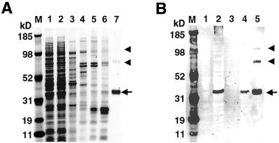
Figure 5.
Expression and purification of the PfPuf1 RNA-binding domain in a bacterial expression system. (A) SDS–PAGE analysis of protein samples. Lane 1, lysate of induced bacteria; lane 2, lysate passed through a Ni–NTA agarose column; lane 3, 15 mM imidazole wash; lane 4, 30 mM imidazole wash; lane 5, 50 mM imidazole wash; lane 6, 80 mM imidazole wash; lane 7, 200 mM imidazole elution. Proteins were electrophoresed in a 4–12% NuPAGE gradient gel and visualized by Coomassie blue staining. (B) An immunoblot of PfPuf1 RNA-binding domain expression. The samples were separated in a 4–12% SDS–PAGE gel and transferred to nitrocellulose membrane for immunoblotting with monoclonal anti-His tag antibody. Lane 1, lysate of uninduced bacteria; lane 2, lysate of induced bacteria; lane 3, 30 mM imidazole wash; lane 4, 80 mM imidazole wash; lane 5, 200 mM imidazole elution. The 40 kDa PfPuf polypeptide eluted in 200 mM imidazole is indicated by an arrow. Two bands corresponding to the dimers and trimers of the 40 kDa protein are indicated with arrowheads. The MultiMark multi-colored standard (Invitrogen) is indicated in kDa (M).

