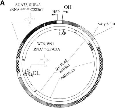
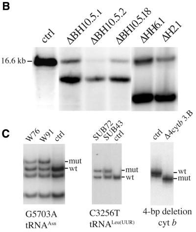
Figure 1.
Characterization of cell lines with mtDNA mutations used in this study. (A) Diagram illustrating the location of two point mutations, two large deletions and a 4 bp deletion in the apocytochrome b gene in the human mtDNA. (B) Southern blot showing the heteroplasmic mtDNA deletions in some of the cell lines used. Total DNA extracted from the different cell lines was digested with PvuII and analyzed by Southern hybridization to a probe corresponding to the ND1 gene. (C) PCR/RFLP analyses of the two pathogenic point mutations and PCR/LP of the apocytochrome b 4 bp deletion (A). The cell lines with tRNA mutations used in this study were heteroplasmic. Cell lines harboring homoplasmic levels of apocytochrome b 4 bp deletion, a 7.5 kb deletion and wild-type genomes were used in the hybrid experiments described below.


