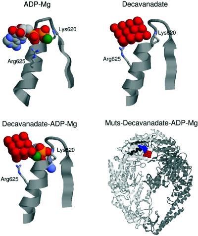
Figure 5.
Resulting structures of docking ADP–Mg, decavanadate and decavanadate–ADP–Mg on the nucleotide-binding domain of E.coli MutS. Docking was carried out as described in Materials and Methods. The three-dimensional structure of the different ligands with the Walker A region complex is shown except the right-hand lower panel where a panoramic view of the MutS dimer–ADP–Mg–decavanadate complex is shown.

