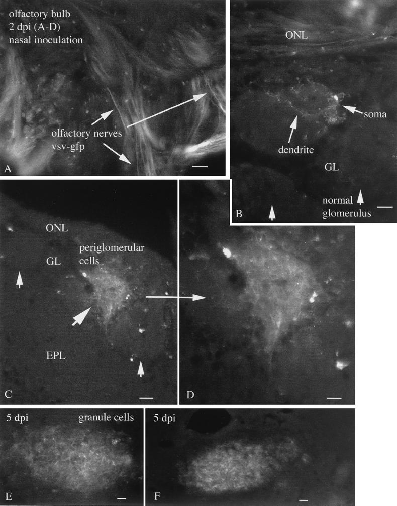
FIG. 1.
Nasal infection 2 to 5 days p.i. (A) Two days after nasal inoculation, VSV-GFP can be found in the olfactory nerves innervating the surface of the olfactory bulb (arrows). Scale bar, 30 μm. (B) A periglomerular cell and its dendrites show GFP fluorescence. Adjacent glomeruli show no sign of infection. Scale bar, 25 μm. (C) A single glomerulus shows strong infection, with many cells expressing GFP. Adjacent glomeruli show only a small amount of infection in one or two cells. Scale bar, 25 μm. (D) Higher magnification of panel C showing dense packing of infected periglomerular cells. Scale bar, 12 μm. (E) Near the center of the bulb, a high level of infection is found. Scale bar, 15 μm. (F) More caudally, a group of cells near the subventricular zone are infected. Scale bar, 15 μm. ONL, olfactory nerve layer; GL, glomerular layer; EPL, external plexiform layer.

